Home

מצטבר סטראוטיפ מערבי radio tibiabin קמעונאות מרגל מקרי

Tibia Fracture | Orthopedic Centers of Colorado

PDF] Tibialization of Fibula in Treatment of Major Bone Gap Defect of the  Tibia: A Case Report | Semantic Scholar
PDF] Tibialization of Fibula in Treatment of Major Bone Gap Defect of the Tibia: A Case Report | Semantic Scholar
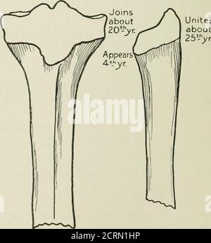
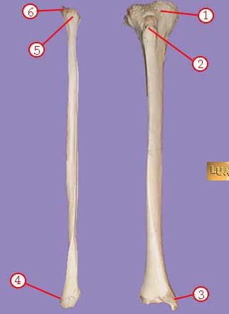
Radiography and radio-therapeutics . Appearsbefore orshortlyafterbirth.  Joins body about20t.h year.. Unites. about 25,-V Lower end of femur. Upper  end of tibia. Upper end of fibula. Fig. 190.—Diagram to show the epiphyses
Radiography and radio-therapeutics . Appearsbefore orshortlyafterbirth. Joins body about20t.h year.. Unites. about 25,-V Lower end of femur. Upper end of tibia. Upper end of fibula. Fig. 190.—Diagram to show the epiphyses
Fibula, or calf bone

Tibia, or shinbone
Tibia, or shinbone

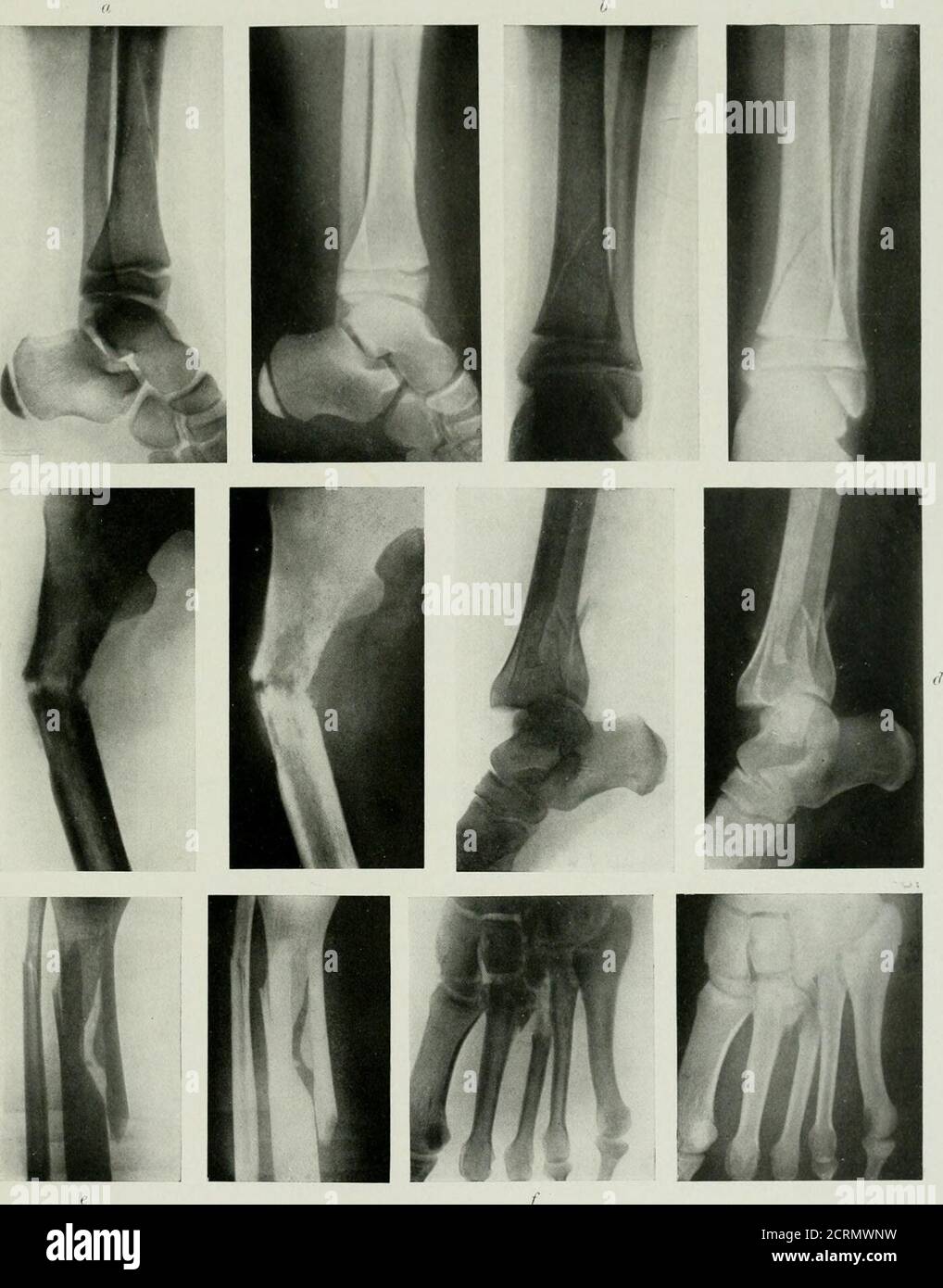
Radiography and radio-therapeutics . Fig. 225.—Fracture of os calcis  (gunshot wound).. PLATE XXXVI. — FiJACTrRKS of Lkg, Ankle, axu Foot. «,  Oblique fracture of shaft of tibia, lateral view, shows epiphyses
Radiography and radio-therapeutics . Fig. 225.—Fracture of os calcis (gunshot wound).. PLATE XXXVI. — FiJACTrRKS of Lkg, Ankle, axu Foot. «, Oblique fracture of shaft of tibia, lateral view, shows epiphyses

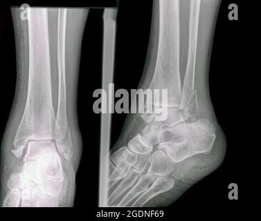
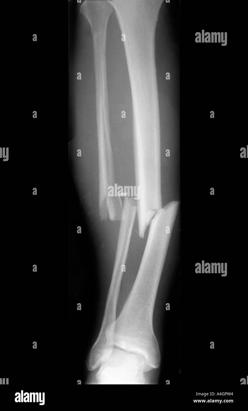
Fracture of the distal tibia and fibula. X-ray of a 57 year old male Stock  Photo - Alamy
Fracture of the distal tibia and fibula. X-ray of a 57 year old male Stock Photo - Alamy

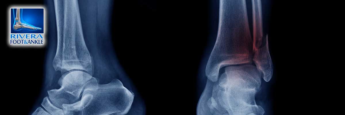
Rivera Foot and Ankle - Podiatrist Houston

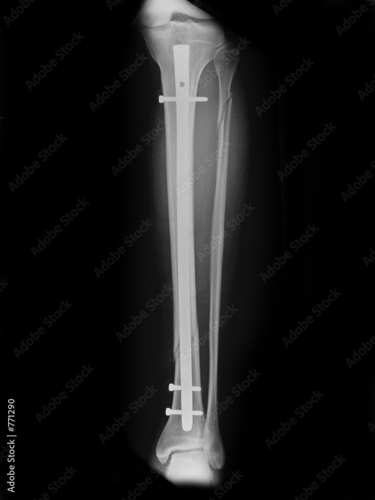
radio fracture de jambe enclouage Stock Photo | Adobe Stock

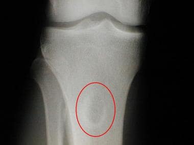
Brodie abscess | Radiology Reference Article | Radiopaedia.org

JCM | Free Full-Text | Benign Bone Tumors: An Overview of What We Know Today

Metaphysis - Wikipedia

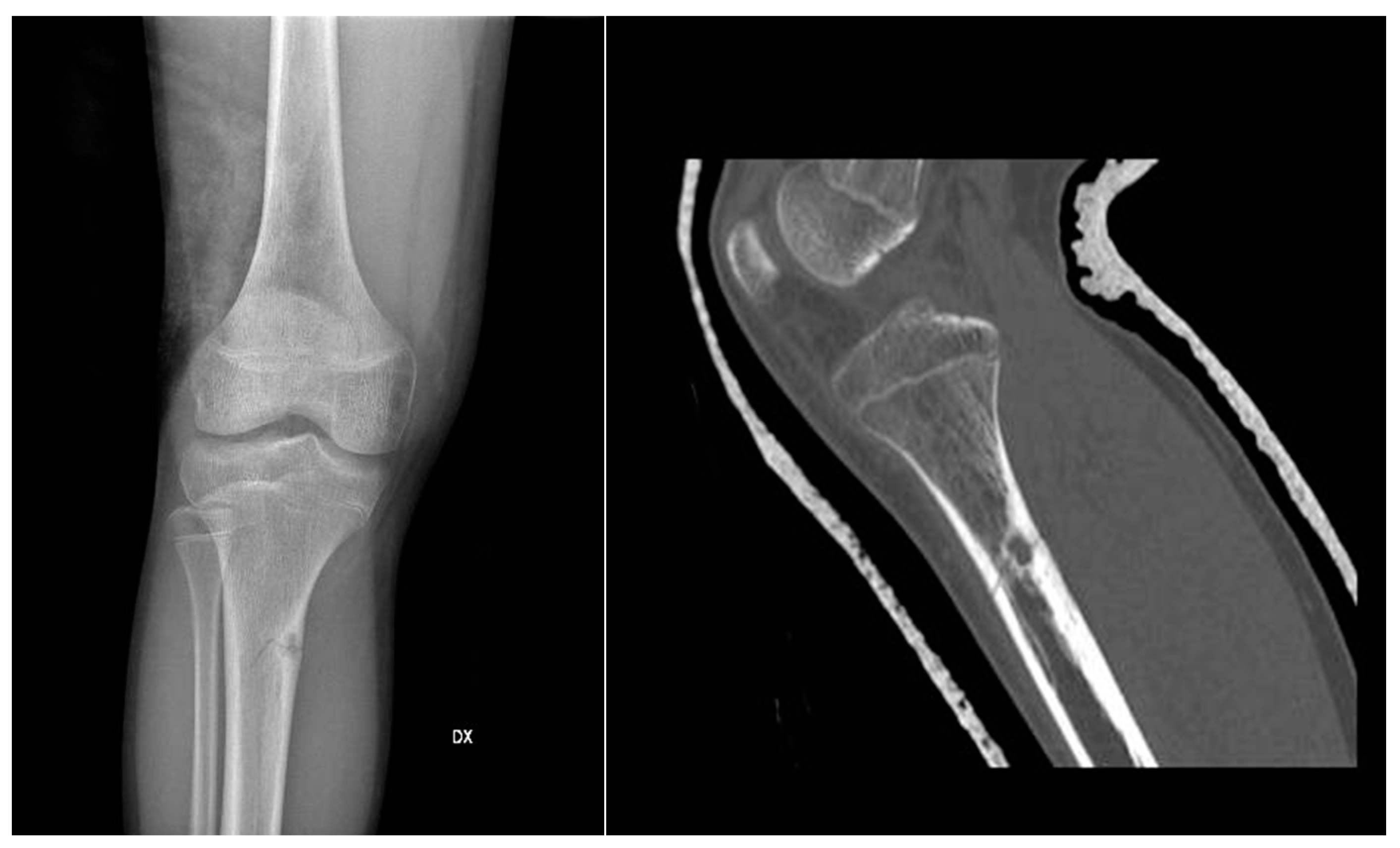
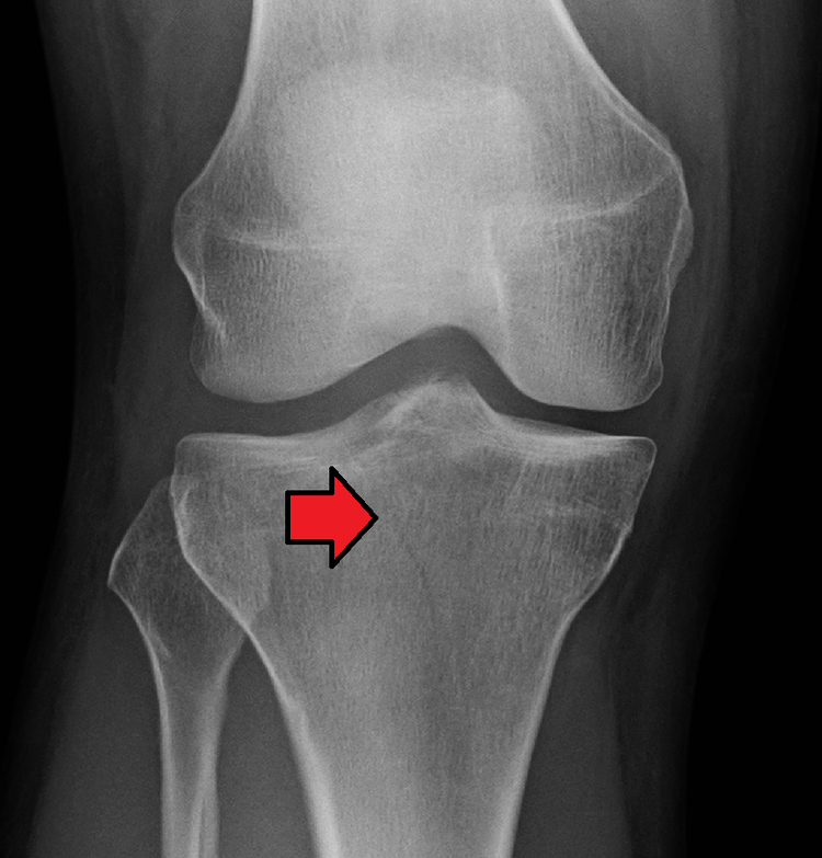
Chondromyxoid Fibroma Imaging: Practice Essentials, Radiography, Computed  Tomography
Chondromyxoid Fibroma Imaging: Practice Essentials, Radiography, Computed Tomography

Tibia, or shinbone
Tibia, or shinbone

Radio : fracture tibia + fibula | Sur un chiot, avant réduct… | Flickr

X-ray Image Of Broken Leg. AP View, Shows Tibia And Tibula Fracture. Stock  Photo, Picture and Royalty Free Image. Image 40036657.
X-ray Image Of Broken Leg. AP View, Shows Tibia And Tibula Fracture. Stock Photo, Picture and Royalty Free Image. Image 40036657.

Osteochondroma | Radiology Reference Article | Radiopaedia.org

Full-length AP and lateral radiographs of the tibia and fibula showing... |  Download Scientific Diagram
Full-length AP and lateral radiographs of the tibia and fibula showing... | Download Scientific Diagram

Measurement values for the bone morphologies of the femur and tibia.... |  Download Scientific Diagram
Measurement values for the bone morphologies of the femur and tibia.... | Download Scientific Diagram

Fracture of the Proximal Tibia

New Page 1
New Page 1

Premium Photo | X-ray foot normal joint and osteochondroma of distal tibia  cause pressure effect to the fibula
Premium Photo | X-ray foot normal joint and osteochondroma of distal tibia cause pressure effect to the fibula

Incomplete fracture | Radiology Reference Article | Radiopaedia.org

Fractured tibia hi-res stock photography and images - Alamy

Target INI CET MD/MS - #GIANT_CELL_TUMOR #RADIO #ORTHO GCT are: · Age  group-20–40 years · Location: Distal femur, proximal tibia, Distal radius,  Proximal humerus, Sacrum, ilium · Features: A. Most lesions begin
Target INI CET MD/MS - #GIANT_CELL_TUMOR #RADIO #ORTHO GCT are: · Age group-20–40 years · Location: Distal femur, proximal tibia, Distal radius, Proximal humerus, Sacrum, ilium · Features: A. Most lesions begin